Student’s Name
Professor’s Name
Course
Date
Circulatory System of a Teleost Fish
Introduction
Teleost fish are the most advanced species of fish and are dominant in both marine and fresh water habitat. They are found in turbulent waters, especially deep oceans and rivers. The body shape of teleost fish depends on the habitat. The fish body is optimized for low resistance when swimming by ensuring streamline movement through water. The most identical part of the fish is its skeleton and tail referred to as caudal fin.
General Layout and the Function of Circulatory System of a Teleost Fish
The general layout of circulatory system consists of a single circuit in which blood is pumped to other organs by the heart. Blood flows through a series of blood vessels namely, arteries, arterioles, capillaries and veins. The circulatory system also forms a passage for other requirements like respiratory gases and nutrients. Blood forms the medium for transporting nutrients and respiratory gas to and from the body tissues (Ramel).
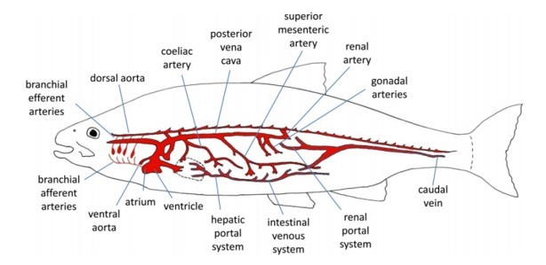
Figure 1. Fish circulation layout ("PG Certificate in Sustainable Aquaculture")
The Heart
Heart of teleost fish has four chambers connected in series from sinus venosus, atrium, ventricle and bulbous arteriosus. The heart is enclosed within a pericardial cavity which is semi rigid in nature. Flow of blood starts from venous where the blood enters the ventricle through sinus venous. Along the flow of blood to the atrium, back flow is prevented by the Sinoatrial valves. The atrium has a thin muscular wall which allows contraction and relaxation of muscles. The contraction propels the blood through atrio-ventricular valve into the ventricle. Heart ventricle acts as the pumping site in the heart where blood gathers pressure for flowing throughout the body. The ventricle is filled by contraction of atrium and also directs inflow of blood from central veins especially in the diastole phase.
Once the blood is in the ventricle, the ventricle pumps blood through bulbous arteriosus into highly compliant bulbous arteriosus into ventral aorta. Back flow of blood to the ventricle is prevented by a series of semilunar valves in the bulbous arteriosus. Once the blood is pumped out of the ventricle, the ventricular muscle begins to relax ready for another diastole action. Blood to the heart muscles is blood by coronary artery which is attached to the ventral aorta.
Cardiac Cycle
The heart has four chambers which must be activated sequentially for a smooth circulation of blood. Muscular layers are needed in different sections of each chamber to affect a smooth synchronized activity. Cardiac cycle is initiated by the pace maker cells which create electrical wave that spreads throughout the heart. The electrical waves are recorded as electrocardiogram which represents the heart beat or heart rate. Cardiac output represents the volume of blood that is pumped by the heart per unit time. For most fish, cardiac output are affected by the changes I stroke volume rather than the heartbeat.
The cardiac cycle starts in the sinus venous and atrium when the electric wave is activated. The wave causes contraction of the muscles in the atrial wall giving rise in pressure hence blood flows into ventricle through atrio-ventricular. The blood flow increases the pressure in ventricle and expands the ventricular wall. Once the wall is under pressure, an electrical activity occurs in the ventricle causing contraction of the muscle increasing pressure of blood in the ventricle. Increasing pressure results into closure of atrio-ventricular valves which prevent back flow of blood into the atrium. High pressure in the ventricle results to an increase in ventral aorta pressure while back flow is prevented by the semi lunar valves in bulbous arteriosus. Pressure in ventral aorta declines slowly as blood flows to the gills and other organs (Ramel). The cycle is completed and another cycle is initiated when blood enters the atrium.
Blood Circulation
The primary blood circulation is connecting the heart and the gills in series. The ventral aorta splits into four pairs of branchial arteries that supply the gills with blood. There are two major routes in the gills, first is the blood flow over venous compartment. The second path is where blood leaves gills by efferent branchial arteries to supply dorsal aorta and head through carotid arteries. Secondary circulation is what differentiates the teleost form other fish. The network of arteries supplies blood to the skin and internal surfaces. Secondary veins then drain blood into vein of primary circulation.
Conclusion
A fish’s heart has four chambers but unlike human beings, the heart is not muscular. Therefore, the chambers are located one behind the other. The first chamber is called the sinus venosus, second atrium, third ventricle and lastly the bulbous arteriosus. Sinus venosus is the collecting chamber which collects blood from lateral veins, anterior cardinals and posterior cardinals. Atrium receives blood from sinus venous and pushes the blood into the ventricle. Ventricle is the only muscled chamber and builds up pressure to allow blood flow around the body. Bulbous arteriosus chamber is primarily elastic and works to reduce pulsed nature of blood giving it a more even and constant flow.
Works Cited
“PG Certificate in Sustainable Aquaculture.” The Fish Site, n.d., http://www.thefishsite.com/learn/modular_course_structure.php. Accessed 17 May 2017.
Ramel, Gordon. "The Earth Life Web, The Circulatory System Of Fish". Earthlife.Net, 2017, http://www.earthlife.net/fish/blood.html. Accessed 17 May 2017.